FMF Certification
Preeclampsia screening
Preeclampsia (PE) is an important cause of maternal and perinatal mortality and morbidity. Consequently, a major challenge in modern obstetrics is early identification of pregnancies at high-risk of preterm PE and undertaking of the necessary measures to improve placentation and reduce the prevalence of the disease. There is now evidence that a combination of maternal demographic characteristics, including medical and obstetric history, uterine artery pulsatility index (PI), mean arterial pressure (MAP) and placental growth factor (PlGF) at 11-13 weeks' gestation can identify a high proportion of pregnancies at high-risk for preterm PE. Such early identification of the high-risk group for preterm PE is important because the risk is substantially reduced by the prophylactic use of low-dose aspirin starting from 11-13 weeks.
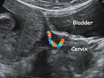
Color Doppler of uterine arteries
Requirements for certification
Please attend all online courses through our new FMF website.
From March 2026, registration will be required to qualify for a license extension for the risk calculation software. All new licenses will be issued exclusively through the new website.
To apply for a new license or request a license extension, please follow instructions published on your original FMF Page. Please continue using your original FMF page on this website to apply for and access compatible license files via the Software section.
Note: Some countries have additional certification requirements.
USA: Visit www.fetalmedicineusa.com for NT and PE accreditation instructions and contact details.
Australia: Contact RANZCOG for accreditation and image submission details.
Further updates and requirements will be posted online soon.
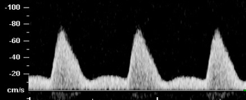
Uterine artery waveform
Protocol for measurement of the uterine artery PI
- The gestational age must be between 11 weeks and 13 weeks and six days.
- Sagittal section of the uterus must be obtained and the cervical canal and internal cervical os identified. Subsequently, the transducer must be gently tilted from side to side and then colour flow mapping should be used to identify each uterine artery along the side of the cervix and uterus at the level of the internal os.
- Pulsed wave Doppler should be used with the sampling gate set at 2 mm to cover the whole vessel and ensuring that the angle of insonation is less than 30º. When three similar consecutive waveforms are obtained the PI must be measured and the mean PI of the left and right arteries be calculated.